- Research
- Open access
- Published:
Dependence on visual information in patients with ACL injury for multi-joint coordination during single-leg squats: a case control study
BMC Sports Science, Medicine and Rehabilitation volume 16, Article number: 87 (2024)
Abstract
Background
The influence of vision on multi-joint control during dynamic tasks in anterior cruciate ligament (ACL) deficient patients is unknown. Thus, the purpose of this study was to establish a new method for quantifying neuromuscular control by focusing on the variability of multi-joint movement under conditions with different visual information and to determine the cutoff for potential biomarkers of injury risk in ACL deficient individuals.
Methods
Twenty-three ACL deficient patients and 23 healthy subjects participated in this study. They performed single-leg squats under two different conditions: open eyes (OE) and closed eyes (CE). Multi-joint coordination was calculated with the coupling angle of hip flexion, hip abduction and knee flexion. Non-linear analyses were performed on the coupling angle. Dependence on vision was compared between groups by calculating the CE/OE index for each variable. Cutoff values were calculated using ROC curves with ACL injury as the dependent variable and significant variables as independent variables.
Results
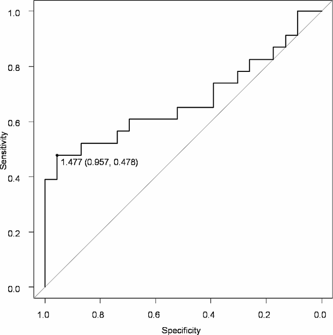
The sample entropy of the coupling angle was increased in all groups under the CE condition (P < 0.001). The CE/OE index of coupling angle variability during the descending phase was higher in ACL deficient limbs than in the limbs of healthy participants (P = 0.036). The CE/OE index of sample entropy was higher in the uninjured limbs of ACL deficient patients than in the limbs of healthy participants (P = 0.027). The cutoff value of the CE/OE index of sample entropy was calculated to be 1.477 (Sensitivity 0.957, specificity 0.478).
Conclusion
ACL deficient patients depended on vision to control multiple joint movements not only on the ACL deficient side but also on the uninjured side during single leg squat task. These findings underscore the importance of considering visual dependence in the assessment and rehabilitation of neuromuscular control in ACL deficient individuals.
Background
Anterior cruciate ligament (ACL) injury is the one of the most common injury of the knee in sports [1]. The incidence rate of ACL injuries remained relatively stable between 1990 and 2010, especially in females [2]. Moreover, patients who have undergone ACL reconstruction often require revision or suffer an ACL injury on the contralateral side. In the first 5 years after ACL reconstruction, the rate of new ACL injury is higher than the rate of primary ACL injury in the general population [3]. These high ACL injury rates may be due in part to the lack of effective prevention programs before injury and after ACL reconstruction.
Noncontact mechanisms account for 70–76% of all ACL injuries [4] and occur most commonly during dynamic activities involving rapid deceleration and landing [5, 6]. Performing these movements with less risk of ACL injury requires more skillful neuromuscular control. In previous studies, neuromuscular control systems have often been represented by the movement of the centre of pressure (COP). Fernandes TL et al. observed that during single-leg standing and squat tasks, athletes with ACL injuries exhibited greater lateral shifts in the COP than did healthy subjects [7]. Similarly, Nematollahi M et al. reported that the trembling component of the COP, which reflects peripheral systems such as muscle activity, was significantly greater in individuals with ACL deficiency under both single-leg and double-leg conditions, indicating increased instability [8]. Bodkin SG et al. reported no significant difference in the average velocity of the COP during one-legged stance postural control between patients who had undergone ACL reconstruction and healthy subjects, suggesting that ACL reconstruction may restore some aspects of postural control to preinjury levels [9]. However, Steffen K et al. found no correlation between COP movement velocities, both anterior-posterior and lateral, during static and dynamic postural control and the risk of ACL injury in female elite handball and soccer players [10]. This indicates that poor movement specific to those at high risk of ACL injury is inadequately measured by COP and that it is difficult to identify features of the neuromuscular control system. Moreover, several studies suggest that noncontact ACL injury results from multi-plane joint moment caused by multi-directional ground reaction forces [5, 11, 12]. These studies show that controlling joint motion across multiple joints could lead to postural stabilization and prevention of ACL injury. The nonlinear analyses of motion variability associated with ACL injury or reconstruction have focused on two different joints or two joint motions [13,14,15]. On the other hand, it has been shown that more than three joint motions, including others in the knee joint, may be involved in the risk of noncontact ACL injury. Video analysis at the time of ACL injury reported low hip flexion angles [16], and weakness of the hip abductor muscle strength was a risk factor for non-contact ACL injuries [17]. In addition, a decrease in absorption in the lower extremity due to a smaller hip flexion angle motion [18], this is an energy absorption strategy that relies on distal joints such as the ankle joint and may increase the knee valgus motion [19]. It is speculated that the combined occurrence of these factors increases the risk of ACL injury. These joint movements can be controlled by muscles, unlike knee valgus motion which does not have a primary active muscle, so skillful control of these joint movements may be useful in preventing ACL injury.
Recent studies have shown that coordination patterns change depending on the availability of visual information [20]. Studies of stability control related to ACL injury have examined the influence of vision in static assessments such as quiet standing or one-legged standing. In several previous reports, COP deviation in patients after ACL reconstruction was greater with closed eyes than with open eyes, and these values showed a greater range of elevation than did those on the healthy or uninjured side [21,22,23].. However, prevention of ACL injuries or revision after ACL reconstruction requires stable postural control in more dynamic situations. Trulsson et al. reported deviations in muscular activity between the injured and noninjured sides in individuals with ACL injuries during single-leg squats, suggesting altered sensorimotor control [24]. Therefore, the influence of vision on the neuromuscular control system in more complex tasks, such as the single-leg squat, should be considered.
The purpose of this study was to reveal differences in neuromuscular control in ACL-deficient patients during single-leg squats with different visual information via nonlinear analysis for multiple joint movements. We hypothesized that ACL-deficient patients would exhibit more variability during movement in both of injured side and uninjured side, and that reduced visual information would further manifest their characteristic movements variability.
Methods
Participants
This study was approved by the Ethical Review Committee for Medical Research Involving Human Subjects in accordance with the Declaration of Hiroshima University (ID number: C-274-1). Twenty-three patients with non-contact ACL injury (23 affected knees) aged from 16 to 42 years (11 males and 12 females; mean age, 21.7 ± 6.9 years old) participated in this study. The recruitment period for this study was between July 2019 and June 2022. The inclusion criteria were as follows: outpatients of the Department of Orthopaedic Surgery, Hiroshima University Hospital, who completed junior high school or other courses, diagnosed by an experienced orthopaedic surgeon as having ACL injury based on MRI imaging findings and physical findings, requiring ACL reconstruction, and able to walk alone. The surgeon was trained to uniformly evaluate the physical examination findings, including the pivot shift test and the Lachman test. Patients were excluded if they had any of the following: under 16years old, bilateral ACL injury, history of lower limb injuries within 2 years, ligament reconstruction within 2 years, knee or hip joint arthroplasty or high tibial osteotomy, neuromuscular disorder, history of stroke or cardiovascular disease, or any other gait abnormalities. For comparison, 23 healthy subjects matching age and body size with no history of neuromuscular disorder or orthopaedic problems in the lower limbs participated. Participant characteristics are shown in Table 1. All participants in this study gave informed consent using documentation and signed a consent form.
A prior power analysis for sample size was performed with G*Power (version 3.1; Franz Faul, Kiel University, Kiel, Germany); for an effect size of 0.3, power of 0.80, an α level of 0.05, and numerator degrees freedom of 1 and 2, number of groups of 6; a total of 90 and 111 samples for main effects, and 111 samples for interactions were needed, respectively. Therefore, there was a minimum of 23 samples for each condition and for each group considering possible dropout in this study.
Procedure
Kinematic data on the patient motion were acquired using a three-dimensional motion analysis system (VICON NEXUS; Vicon Motion Systems, Oxford, UK) with 16 infrared cameras (Vicon Motion Systems, Oxford, UK) operating at 200 Hz. Before each measurement session, devices were calibrated, and the mean calibration residuals for trials were under 1.00 mm.
Infrared-reflecting markers 14 mm in diameter were attached to 45 landmarks including the left front head, right front head, left back head, right back head, 7th cervical vertebrae, 10th thoracic vertebrae, clavicle, sternum, right back, bilaterally acromion, lateral epicondyle approximating the elbow joint, wrist bar thumb side, and pinkie side, head of the 2nd metacarpus, anterior superior iliac spine, posterior superior iliac spine, great trochanter, lateral aspects of the thighs, lateral and medial epicondyles of the femur, lateral aspects of the shanks, lateral and medial condyles of the tibia, lateral and medial malleoli, head of the 2nd metatarsal heads, and the calcaneal tuberosity. Motion trials were captured as the participant performed single-leg squats (SLSs). Participants performed the actual task after completing a minimal set of fewer than five consecutive SLSs with their eyes open as a preliminary exercise. Participants were instructed to perform 12 SLSs with their hands on hips; requirements for the flexion angles of the joints and the depth of the squat were not specified. The SLSs were conducted in sync with a metronome set at 120 bpm, such that the metronome emitted a clicking sound once at the lowest position of the squat and once at the highest position. Participants performed the exercises under two randomized order conditions; the eyes-opened (OE) and eyes-closed (CE), with the supporting leg, which was defined as the lower limb on the supporting side when kicking a ball, in healthy subjects (Healthy) and both the ACL-deficient (ACLD) side and contralateral uninjured side (Uninjured) in patients with ACL injury. Successful trials were those in which the participants performed 12 repetitions without the opposite lower limb touching the ground and performed in rhythm. The first and last one each were excluded, and the 10 SLSs were analysed.
Data processing
The lower limb joint angles and centre of mass (COM) were calculated using the processing software Body-Builder (Vicon Motion Systems, Oxford, UK) based on collected marker coordinates. The centre of a participant’s ankle joint was estimated as the midpoint between the malleoli, while the knee joint centre was estimated as the midpoint between the lateral and medial epicondyles of the femur and the lateral and medial condyles of the tibia. The hip joint centre was estimated based on a previous study [25]. The collected marker coordinates were used to define the respective local coordinate systems of the fifteen-point body link model consisting of the head, thorax, both upper arms, both lower arms, hands, pelvis, both thighs, both shanks, and both feet. The position of the centre of mass position for each segment was calculated based on body inertia characteristics in a report by Okada et al. [26], and all composite centres of mass for all segments were used as the whole-body COM. A single squat was identified as the combined descending and ascending phases of a SLS indicated by the COM vertical displacement between the vertical maximum position.
Multiple joint coordination
Hip flexion-extension, hip abduction-adduction, and knee flexion-extension motions are associated with ACL injury [6]. The coordination of these three joint motions, hip flexion (+)–extension (-), hip abduction (+)–adduction (-) and knee flexion (+)–extension (-), and the coupling angle (CA) were obtained from the Appendix.
The COM data were divided into ascending and descending phases, and the coupling angle variability (CAV) was calculated for each phase by the Appendix, and the average of 10 SLSs. The sample entropy (SaEn) of the CA was calculated with embedding dimension, and tolerance was set to 2 and 0.2 × SD, respectively [27]. Non-linear analysis processing was conducted in open-source Python (version 3.9) under Jupyter Notebook with pandas, nolds, numpy, sklearn and Anaconda libraries.
Dependence of visual information
To examine the effect of visual acuity, the CE/OE index was calculated for each variable by dividing the CE value by the OE value. Values close to 1 suggest a minimal influence of visual information on balance, while values greater than 1 indicate a greater dependency on vision.
Statistical analysis
The statistical analyses were performed using IBM SPSS 25.0 (SPSS Inc., Chicago, IL, USA). Differences in physical characteristics between groups in the participants were tested with an unpaired t-test. A two-way factorial analysis of variance (ANOVA) was performed to assess the effects of group (Healthy vs. ACLD vs. Uninjured) and condition (eyes-open and eyes-closed) on COP values, CAV and SaEn. One-way ANOVA was performed for the CE/OE index for each variable. All variables are presented as the mean and SD. If significant main effects or interactions were identified using ANOVA, post hoc pairwise comparisons using the Tukey‒Kramer multiple comparisons test were then performed.
Finally, the capacities of dependence on visual information indicators for predicting ACL-injured risk were compared via area under the receiver-operating characteristics (ROC) curves (AUC) analysis. We analysed variables significantly different from healthy subjects for the uninjured side limb of ACL-injured individuals, rather than the injured leg, in order to identify the potential risk of ACL injury. The cutoff value was defined as the point at which the Youden Index of the ROC curve was the largest. All p values were two–sided and p < 0.05 was considered statistically significant.
Results
Effects of ACL injury and condition on CAV and SaEn
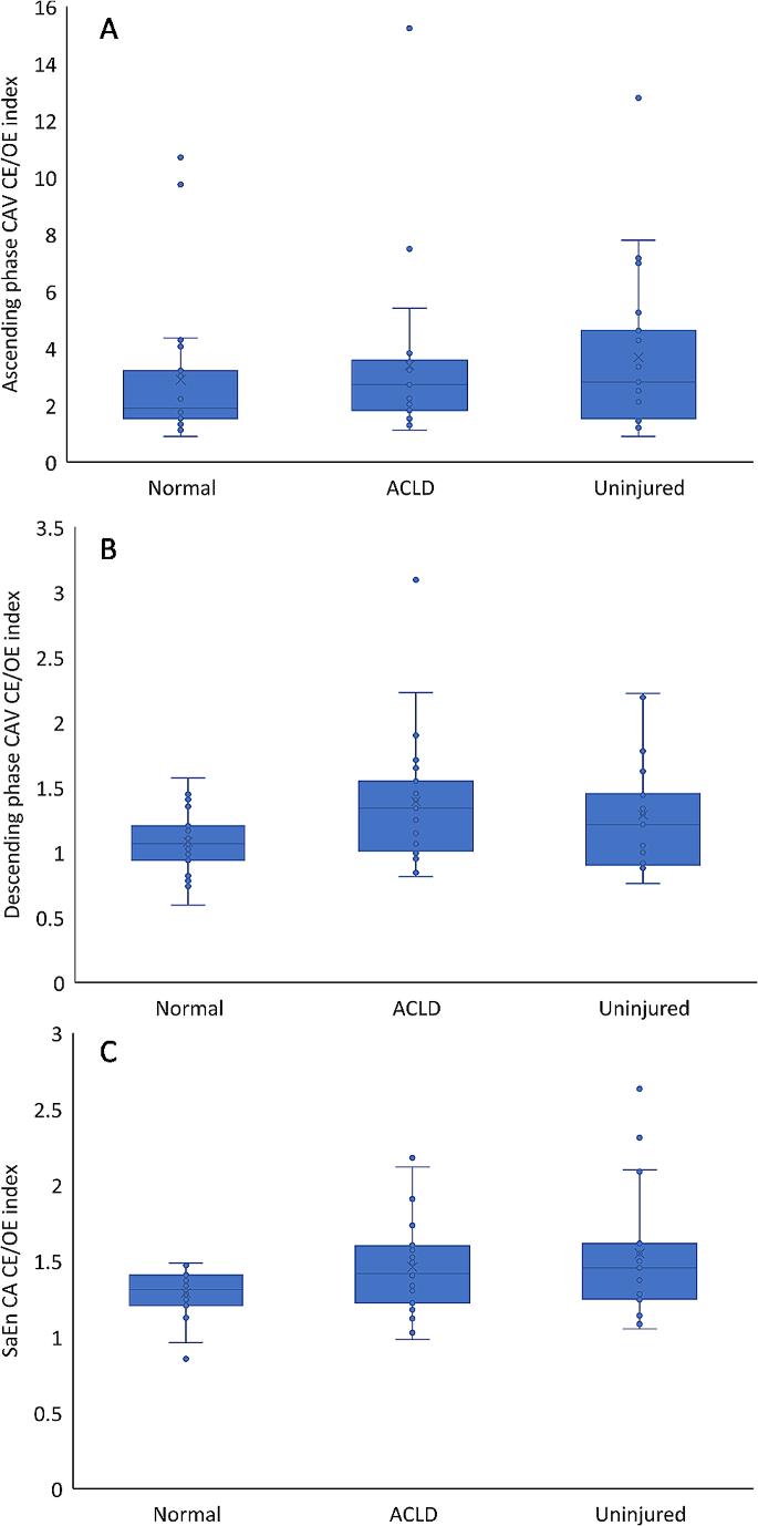
Significant condition-specific effects were observed for ascending-phase CAV (p < 0.001, F = 33.86), descending-phase CAV (p = 0.048, F = 3.99) and SaEn CA (p < 0.001, F = 52.02). Conversely, the main effect of group failed to reach statistical significance. All results of two-way ANOVA are shown in Table 2.
Effects of visual information for CAV and SaEn
Group mean ± 95% confidence intervals, along with individual participant mean outcome measures are presented in Fig. 1, with full statistical analysis reported in Table 3. The CE/OE index of CAV during the descending phase in the ACLD was higher than that in healthy participants (95% CI 0.017-0.60; P = 0.036; Table 3). The CE/OE index of SaEn on the uninjured side was higher than that of healthy participants (95% CI 0.031–0.49; P = 0.027; Table 3.)
ROC curve analysis
ROC curve analysis by CE/OE index of SaEn is shown in Fig. 2. The cutoff of the CE/OE index of SaEn was calculated to be 1.477 (sensitivity 0.957, specificity 0.478), and AUC was 0.677(95% CI 0.513–0.84).
Discussion
The most important finding in this study is that patients with ACL injuries have contralateral neuromuscular control system dysfunction, indicating that stable and continuous joint motion is difficult at multiple joints. These results may quantify the potential risk of ACL injury in individuals with current ACL injuries and may be useful for preventing not only future reinjury of the reconstructed ACL but also new injury of the contralateral ACL.
Our study results showed that, in both healthy subjects and those with ACL injuries, the variability in postural control during SLSs was greater when the subjects’ eyes were closed than when they were open. Specifically, we observed an average increase in variability of 136.8% during the ascending phase and 117.3% during the descending phase, as measured by the CAV, and an average increase of 141.8% in the SaEn CA. This finding indicates greater variability in postural control during dynamic motor tasks with eyes closed. However, there was no difference in variability under both the open-eyes and closed-eyes conditions between the two groups. These results are consistent with the finding of Dingenen et al., who demonstrated no significant difference in single-leg stance COP stability among healthy, ACL-injured, and contralateral limbs under both open-eyes and closed-eyes conditions [28]. In contrast to our findings, prior studies have reported that compared with healthy subjects, individuals with ACL injuries exhibited impaired postural control on not only the injured side but also the contralateral side during static tasks such as static standing and single-leg standing [29, 30]. We speculate that this difference is because we selected the SLS, which is a more dynamic task. Among several dynamic motor tasks, the SLS is more susceptible to dual-task effects [31]. Our study results indicate that it is not appropriate to compare the absolute values of the variability in the movement of SLSs between open and closed eyes. However, several reviews have indicated that a more dynamic assessment is needed for the prevention of noncontact ACL injuries and reinjury [32, 33]. This shows that a novel dynamic postural control assessment index is needed to detect the risk that ACL-injured patients have.
In this study, CE/OE was evaluated to quantify the reliance on visual information in ACL-injured subjects. The results revealed greater variability on the ACLD limb than healthy subjects’ limb during the descending phase of CAV and on the uninjured limbs than healthy subjects’ limb during the entire SaEn. These results suggest that ACL-injured subjects use a postinjury adapted or preinjury potentially visually dependent movement strategy. ACL-injured patients are known to have different motor patterns on the contralateral side compared to healthy subjects [34], and lack of visual information promotes a more rigid movement pattern [35]. This might show that ACL-injured patients exhibit more rigid movement patterns when required to perform the dynamic task of collecting more sensory information due to the loss of visual information. In order to understand the clinical significance of these variables, we used ROC analysis to determine the cutoff risk of ACL injury on the uninjured side limb, which is less susceptible to changes in joint motion due to ACL injury. The cutoff value was 1.477, and the sensitivity was reasonably high, indicating the possibility of screening for ACL injury risk, and although the specificity is low, it is useful for clinically screening individuals for prevention programs. ACL-injured limbs demonstrated lower kinesthesia [36], fewer somatosensory evoked potentials than healthy subjects [37], and a lack of muscle coactivation modulation [38]. ACL-injured individuals might be implementing adaptations to the reduced afferent input at the knee joint due to ACL deficiency that increases afferent joint sensory input information by increasing joint motion variability [39, 40]. In addition to these studies, the lack of visual information for the ACL-injured subjects may also increase variability in other multi-joint movements, including the hip joint that we evaluated, to increase dynamic afferent joint sensory input. Moreover, Diekfuss et al. evaluated altered brain connectivity that may have predisposed athletes to ACL injury and reported that those who went on to experience an ACL injury had decreased functional connectivity between the left primary sensory cortex and right posterior lobe of the cerebellum [41]. These previous reports suggest that ACL-injured patients have different neuromuscular control systems than healthy subjects even before ACL injury, which may have been highlighted by the visual deficits and dynamically unstable motor tasks in the present study. Future research exploring optimal variability in multiple joints will provide a better understanding of ACL injury prevention.
There are three limitations to this study. First, it is not clear when differences in motor control on the uninjured side occur in ACL-injured patients. The subjects with ACL injuries had lower Tegner activity scores than did the healthy subjects, which may indicate that inactivity affects postural control. In addition, it is known that ACL-injured individuals experience a variety of changes due to injury, one of which affects the contralateral lower extremity [42]. To verify this, a prospective cohort study is needed to determine if there are any differences in the contralateral lower extremity in future ACL injury survivors. Second, this study does not fully demonstrate whether ACL deficiency causes differences in movement characteristics on the ACL-injured side. Therefore, it is necessary to clarify the effects of afferent and efferent neuromuscular control system functions, including tests of proprioceptive function and evaluation of muscle activity. To investigate the effects of ACL deficiency, it is necessary to examine whether changes in movement variability occur when knee joint stability is improved through prospective studies after ACL reconstruction. The application of principal component analysis for dimensionality reduction in this study potentially constrained the multidimensional analysis of the data. Consequently, critical insights into the unique joint motion characteristics of individuals with ACL injuries, such as variability in motion during specific time intervals and the interplay between different conditions, might not have been fully captured. To overcome these limitations, future research should include analytical techniques capable of identifying variability across specific periods, such as Statistical Parametric Mapping [43], to provide a more comprehensive understanding of the factors contributing to ACL injury risk.
Conclusion
Subjects with ACL injuries exhibit increased variability and dependence on visual information during SLSs, as indicated by higher CE/OE indices in multiple joint CAV and SaEn than healthy subjects. These findings underscore the importance of considering visual dependence in the assessment and rehabilitation of neuromuscular control in ACL-deficient individuals.
Data availability
The data sets used and/or analyzed in this study are available from the corresponding author if reasonably requested.
Abbreviations
- ACL:
-
Anterior cruciate ligament
- OE:
-
Open eyes
- CE:
-
Closed eyes
- COP:
-
Center of pressure
- BMI:
-
Body mass index
- MRI:
-
Magnetic resonance imaging
- SLS:
-
Single-leg squats
- ACLD:
-
Anterior cruciate ligament deficient
- COM:
-
Centre of mass
- CA:
-
Coupling angle
- CAV:
-
Coupling angle variability
- SaEn:
-
Sample entropy
- AUC:
-
Area under the curve
- ROC:
-
Receiver-operating characteristic
- ANOVA:
-
Analysis of variance
References
Sun Z, Cieszczyk P, Huminska-Lisowska K, Michalowska-Sawczyn M, Yue S. Genetic determinants of the anterior cruciate ligament rupture in Sport: an Up-to-date systematic review. J Hum Kinet. 2023;87:105–17.
Sanders TL, Maradit Kremers H, Bryan AJ, Larson DR, Dahm DL, Levy BA, Stuart MJ, Krych AJ. Incidence of anterior cruciate ligament tears and Reconstruction: a 21-Year Population-based study. Am J Sports Med. 2016;44(6):1502–7.
Wright RW, Magnussen RA, Dunn WR, Spindler KP. Ipsilateral graft and contralateral ACL rupture at five years or more following ACL reconstruction: a systematic review. J Bone Joint Surg Am. 2011;93(12):1159–65.
Agel J, Arendt EA, Bershadsky B. Anterior Cruciate Ligament Injury in National Collegiate Athletic Association Basketball and Soccer: a 13-Year review. Am J Sports Med. 2005;33(4):524–31.
Boden BP, Dean GS, Feagin JA, Garrett WE. Mechanisms of Anterior Cruciate Ligament Injury. Orthopedics. 2000;23(6):573–8.
Myer GD, Ford KR, Di Stasi SL, Foss KD, Micheli LJ, Hewett TE. High knee abduction moments are common risk factors for patellofemoral pain (PFP) and anterior cruciate ligament (ACL) injury in girls: is PFP itself a predictor for subsequent ACL injury? Br J Sports Med. 2015;49(2):118–22.
Fernandes TL, Felix EC, Bessa F, Luna NM, Sugimoto D, Greve JM, Hernandez AJ. Evaluation of static and dynamic balance in athletes with anterior cruciate ligament injury– A controlled study. Clinics. 2016;71(8):425–9.
Nematollahi M, Razeghi M, Tahayori B, Koceja D. The role of anterior cruciate ligament in the control of posture; possible neural contribution. Neurosci Lett. 2017;659:120–3.
Bodkin SG, Slater LV, Norte GE, Goetschius J, Hart JM. ACL reconstructed individuals do not demonstrate deficits in postural control as measured by single-leg balance. Gait Posture. 2018;66:296–9.
Steffen K, Nilstad A, Krosshaug T, Pasanen K, Killingmo A, Bahr R. No association between static and dynamic postural control and ACL injury risk among female elite handball and football players: a prospective study of 838 players. Br J Sports Med. 2017;51(4):253–9.
Quatman CE, Quatman-Yates CC, Hewett TE. A ‘Plane’ explanation of Anterior Cruciate Ligament Injury mechanisms a systematic review. Sports Med. 2010;40(9):729–46.
Serpell BG, Scarvell JM, Ball NB, Smith PN. Mechanisms and risk factors for noncontact ACL injury in age mature athletes who engage in field or court sports a summary of the literature since 1980. J Strength Conditioning Res. 2012;26(11):3160–76.
Moraiti CO, Stergiou N, Vasiliadis HS, Motsis E, Georgoulis A. Anterior cruciate ligament reconstruction results in alterations in gait variability. Gait Posture. 2010;32(2):169–75.
Pollard CD, Stearns KM, Hayes AT, Heiderscheit BC. Altered lower extremity movement variability in female soccer players during side-step cutting after anterior cruciate ligament reconstruction. Am J Sports Med. 2015;43(2):460–5.
Davis K, Williams JL, Sanford BA, Zucker-Levin A. Assessing lower extremity coordination and coordination variability in individuals with anterior cruciate ligament reconstruction during walking. Gait Posture. 2019;67:154–9.
Olsen OE, Myklebust G, Engebretsen L, Bahr R. Injury mechanisms for anterior cruciate ligament injuries in team handball: a systematic video analysis. Am J Sports Med. 2004;32(4):1002–12.
Khayambashi K, Ghoddosi N, Straub RK, Powers CM. Hip muscle strength predicts Noncontact Anterior Cruciate Ligament Injury in male and female athletes: a prospective study. Am J Sports Med. 2016;44(2):355–61.
Decker MJ, Torry MR, Wyland DJ, Sterett WI, Steadman JR. Gender differences in lower extremity kinematics, kinetics and energy absorption during landing. Clin Biomech Elsevier Ltd. 2003;18(7):662–9.
Dadfar M, Soltani M, Novinzad MB, Raahemifar K. Lower extremity energy absorption strategies at different phases during single and double-leg landings with knee valgus in pubertal female athletes. Sci Rep. 2021;11(1):17516.
Busquets A, Ferrer-Uris B, Angulo-Barroso R, Federolf P. Gymnastics experience enhances the development of bipedal-stance multi-segmental coordination and control during proprioceptive reweighting. Front Psychol. 2021;12:661312.
Pahnabi G, Akbari M, Ansari NN, Mardani M, Ahmadi M, Rostami M. Comparison of the postural control between football players following ACL reconstruction and healthy subjects. Med J Islamic Repub Iran 2014, 28(1).
Henriksson M, Ledin T, Good L. Postural control after anterior cruciate ligament reconstruction and functional rehabilitation. Am J Sports Med. 2001;29(3):359–66.
Dauty M, Collon S, Dubois C. Change in posture control after recent knee anterior cruciate ligament reconstruction? Clin Physiol Funct Imaging. 2010;30(3):187–91.
Trulsson A, Miller M, Hansson GA, Gummesson C, Garwicz M. Altered movement patterns and muscular activity during single and double leg squats in individuals with anterior cruciate ligament injury. BMC Musculoskelet Disord. 2015;16:28.
Kurabayashi J, Mochimaru M, Kouchi M. Validation of the estimation methods for the hip joint center. J Soc Biomechanisms. 2003;27(1):29–36.
Okada H, Ae M, Fujii N, Morioka Y. Body segment Inertia properties of Japanese Elderly. Biomechanisms. 1996;13(0):125–39.
Richman JS, Moorman JR. Physiological time-series analysis using approximate entropy and sample entropy. Am J Physiol-Heart C. 2000;278(6):H2039–49.
Dingenen B, Janssens L, Luyckx T, Claes S, Bellemans J, Staes FF. Postural stability during the transition from double-leg stance to single-leg stance in anterior cruciate ligament injured subjects. Clin Biomech (Bristol Avon). 2015;30(3):283–9.
Sayuri Tookuni K, Neto RB, Augusto C, Pereira M, Rúbio D, Souza D, Maria DJ, Greve A, Ayala DA. Comparative Analysis of Postural Control in individuals with and without injuries on knee anterior cruciate ligament. Acta Ortop Bras. 2005;13(3):333–5403.
Culvenor AG, Alexander BC, Clark RA, Collins NJ, Ageberg E, Morris HG, Whitehead TS, Crossley KM. Dynamic single-Leg Postural Control is impaired bilaterally following anterior Cruciate Ligament Reconstruction: implications for Reinjury Risk. J Orthop Sports Phys Ther. 2016;46(5):357–64.
Talarico MK, Lynall RC, Mauntel TC, Wasserman EB, Padua DA, Mihalik JP. Effect of single-Leg Squat speed and depth on dynamic Postural Control under single-Task and Dual-Task paradigms. J Appl Biomech. 2019;35(4):272–9.
Dai B, Cook RF, Meyer EA, Sciascia Y, Hinshaw TJ, Wang C, Zhu Q. The effect of a secondary cognitive task on landing mechanics and jump performance. Sports Biomech. 2018;17(2):192–205.
Alentorn-Geli E, Myer GD, Silvers HJ, Samitier G, Romero D, Lazaro-Haro C, Cugat R. Prevention of non-contact anterior cruciate ligament injuries in soccer players. Part 1: mechanisms of injury and underlying risk factors. Knee Surg Sports Traumatol Arthrosc. 2009;17(7):705–29.
Goerger BM, Marshall SW, Beutler AI, Blackburn JT, Wilckens JH, Padua DA. Anterior cruciate ligament injury alters preinjury lower extremity biomechanics in the injured and uninjured leg: the JUMP-ACL study. Br J Sports Med. 2015;49(3):188–95.
Abe T, Nakamae A, Toriyama M, Hirata K, Adachi N. Effects of limited previously acquired information about falling height on lower limb biomechanics when individuals are landing with limited visual input. Clin Biomech (Bristol Avon). 2022;96:105661.
Shidahara H, Deie M, Niimoto T, Shimada N, Toriyama M, Adachi N, Hirata K, Urabe Y, Ochi M. Prospective study of Kinesthesia after ACL Reconstruction. Int J Sports Med. 2011;32(5):386–92.
Ochi M, Iwasa J, Uchio Y, Adachi N, Sumen Y. The regeneration of sensory neurones in the reconstruction of the anterior cruciate ligament. J Bone Joint Surg Br. 1999;81(5):902–6.
Suarez T, Laudani L, Giombini A, Saraceni VM, Mariani PP, Pigozzi F, Macaluso A. Comparison in joint-position sense and muscle coactivation between anterior cruciate ligament-deficient and healthy individuals. J Sport Rehabil. 2016;25(1):64–9.
Srinivasan D, Tengman E, Hager CK. Increased movement variability in one-leg hops about 20years after treatment of anterior cruciate ligament injury. Clin Biomech (Bristol Avon). 2018;53:37–45.
Heidarnia E, Letafatkar A, Khaleghi-Tazji M, Grooms DR. Comparing the effect of a simulated defender and dual-task on lower limb coordination and variability during a side-cut in basketball players with and without anterior cruciate ligament injury. J Biomech. 2022;133:110965.
Diekfuss JA, Grooms DR, Nissen KS, Schneider DK, Foss KDB, Thomas S, Bonnette S, Dudley JA, Yuan W, Reddington DL, et al. Alterations in knee sensorimotor brain functional connectivity contributes to ACL injury in male high-school football players: a prospective neuroimaging analysis. Braz J Phys Ther. 2020;24(5):415–23.
Konishi Y, Aihara Y, Sakai M, Ogawa G, Fukubayashi T. Gamma loop dysfunction in the quadriceps femoris of patients who underwent anterior cruciate ligament reconstruction remains bilaterally. Scand J Med Sci Sports. 2007;17(4):393–9.
Pataky TC, Robinson MA, Vanrenterghem J, Donnelly CJW. Simultaneously assessing amplitude and temporal effects in biomechanical trajectories using nonlinear registration and statistical nonparametric mapping. J Biomech. 2022;136:111049.
Acknowledgements
We would like to thank Dr. Kunio SHIMIZU at School of Statistical Thinking for helpful analysis of circular statistics on the manuscript.
Funding
This work was supported by JSPS KAKENHI Grant Numbers JP17K18207 and JP 20K19394.
Author information
Authors and Affiliations
Contributions
MT: acquisition and analysis of data, statistical analysis, drafting the article. AN: conception and design, analysis and interpretation of data, drafting the article. TA: acquisition of data. KH: interpretation of data. NA: final approval of the manuscript.
Corresponding author
Ethics declarations
Competing interests
The authors declare no competing interests.
Contributions
MT: acquisition and analysis of data, statistical analysis, drafting the article. AN: conception and design, analysis and interpretation of data, drafting the article. TA: acquisition of data. KH: interpretation of data. NA: final approval of the manuscript.
Ethics approval and consent to participate
All methods were carried out in accordance with the Declaration of Helsinki. This study was approved by the Ethical Review Committee for Medical Research Involving Human Subjects in accordance with the Declaration of Hiroshima University (ID number: C-274-1). Written informed consent to be involved in the experimental procedures was obtained from all the participants before the start of this study.
Consent for publication
NA.
Additional information
Publisher’s Note
Springer Nature remains neutral with regard to jurisdictional claims in published maps and institutional affiliations.
Electronic supplementary material
Below is the link to the electronic supplementary material.
Rights and permissions
Open Access This article is licensed under a Creative Commons Attribution 4.0 International License, which permits use, sharing, adaptation, distribution and reproduction in any medium or format, as long as you give appropriate credit to the original author(s) and the source, provide a link to the Creative Commons licence, and indicate if changes were made. The images or other third party material in this article are included in the article’s Creative Commons licence, unless indicated otherwise in a credit line to the material. If material is not included in the article’s Creative Commons licence and your intended use is not permitted by statutory regulation or exceeds the permitted use, you will need to obtain permission directly from the copyright holder. To view a copy of this licence, visit http://creativecommons.org/licenses/by/4.0/. The Creative Commons Public Domain Dedication waiver (http://creativecommons.org/publicdomain/zero/1.0/) applies to the data made available in this article, unless otherwise stated in a credit line to the data.
About this article
Cite this article
Toriyama, M., Nakamae, A., Abe, T. et al. Dependence on visual information in patients with ACL injury for multi-joint coordination during single-leg squats: a case control study. BMC Sports Sci Med Rehabil 16, 87 (2024). https://doi.org/10.1186/s13102-024-00875-9
Received:
Accepted:
Published:
DOI: https://doi.org/10.1186/s13102-024-00875-9

